The High vs. Low PEEP Debate in Mechanical Ventilation: Are We Just Blowing Hot Air?

In the world of mechanical ventilation, few topics spark as much debate—and as many eye rolls—as the discussion around PEEP settings. Yes, that’s right, we’re talking about Positive End-Expiratory Pressure, the setting that keeps our patients’ lungs open, their oxygen levels up, and our Journal and conferences locked in never-ending debates. But while some clinicians treat this debate like it’s a life-and-death matter (because, well, it kind of is), let’s take a moment to breathe deeply and look at the High vs. Low PEEP controversy with a bit of humor. After all, who says we can’t laugh at ourselves since we really don’t know

The High PEEP Advocates: More is More, Right?

If you’ve ever met a High PEEP enthusiast, you know they’re a passionate bunch. For them, cranking up the PEEP is akin to turning up the volume on your favorite song—louder is always better. They argue that higher PEEP levels keep the alveoli open, improve oxygenation, and prevent the dreaded atelectasis from rearing its ugly head. And, in many cases, they’re right. For patients with severe ARDS, a higher PEEP can indeed make the difference between life and death.

But let’s be honest—High PEEP proponents can sometimes take it a bit too far. It’s as if they believe that more pressure will magically solve every problem, much like the guy at the gym who thinks adding more weight to the bar will instantly turn him into the Hulk. But here’s the thing: while High PEEP can be a powerful tool, it’s not a one-size-fits-all solution. There are risks, like barotrauma and hypotension, that can make this approach a double-edged sword. So, while we applaud their enthusiasm, maybe it’s time to dial it back just a notch. Not every patient needs to feel like they’re in a wind tunnel.

The Low PEEP Loyalists: Less is More (And Also Safer)

On the other side of the battlefield, we have the Low PEEP loyalists—those who believe that when it comes to PEEP, less is definitely more. For them, the idea of pumping high pressures into delicate lung tissue is as appealing as putting pineapple on pizza (a practice that sparks its own set of heated debates: The Hawaiians ruined the Pizza according to Anger from Inside Out)).

Low PEEP advocates argue that keeping PEEP low reduces the risk of barotrauma, minimizes the potential for hemodynamic instability, and generally makes life easier for the lungs. And they’ve got sorta a little point.

Low PEEP settings are often preferred for patients with conditions like COPD or when there’s a concern about blood pressure dropping faster than a Wi-Fi signal during a Zoom meeting. The Low PEEP approach is all about caution, careful monitoring, and avoiding the pitfalls of too much pressure. It’s the respiratory equivalent of the “minimalist” movement—why add more when less will do just fine?

The Gray Area: Why the Debate is Far From Over

Of course, as with most things in life, the truth lies somewhere in the middle. The High vs. Low PEEP debate isn’t black and white—it’s more like fifty shades of gray, each with its own clinical nuances. Patient variability, underlying conditions, and real-time monitoring mean that what works for one patient might not work for another. It’s like trying to find the perfect temperature for your shower—everyone’s got their own sweet spot.

But wait, how about the science, the prospective studies, the meta analysis, the Biblical societies guidelines, the PEEP-FiO2 tables. Those solved the problem and ended the debate right ? I’ll leave you to laugh at that for a little bit…..

The reality is that the High vs. Low PEEP debate is as much about philosophy as it is about physiology. Some clinicians are risk-takers, willing to push boundaries for the sake of better oxygenation. Others prefer a more conservative approach, focusing on steady, reliable outcomes. And both sides have valid points. So, while we may never fully resolve the debate, it’s important to keep the conversation going. Because in the end, what really matters is doing what’s best for the patient in front of us—even if it means admitting that maybe, just maybe, we’re all just blowing a little hot air.

Should we automate PEEP ?

Before we dig in this tough topic, let’s agree or disagree that that there is nothing called optimal or best PEEP. Regardless of the PEEP level used, there will be overdistention in some areas, some under distention and collapse in other areas of the lungs.

Here are some points to keep in mind when we talk PEEP

  • The normal and the diseased lungs are markedly heterogenic with each lobule or even each of the approximately 500 million alveoli have their own mechanics
  • The pleural pressures are not uniform and thus the trans-alveolar pressures will vary depending on the location of the alveoli
  • Not all Lungs especially ARDS are recruitable
  • “Not all ARDS are the same” but studies don’t differentiate ARDS phenotypes
  • “PEEP does not recruit, rather prevents derecruitment”
  • We don’t have much agreements in the literature on how to set PEEP or if PEEP affect mortality
  • The respiratory mechanics change frequently, even between breaths to breaths but we don’t change the PEEP frequently enough, so what was good 10 minutes ago might not be good now

Why automate PEEP or let the machine adjust PEEP?

Simple answer, because we honestly don’t know how despite 50 years of research, and we can’t do it continuously or be at the bedside all the time while the respiratory mechanics are continuously changing

What are the benefits?

  • To minimize the over and under distention of one level of PEEP
  • The ventilator can be consistent on the way it adjust the PEEP per specific algorithms

Are there any modes that currently automate PEEP?

Yes, INTELLiVENT‑ASV from Hamilton was the first mode to automate PEEP, among all other parameters of ventilation and proved to be an effective mode of ventilation from intubation to extubation. However the algorithms used is the ARDSnet PEEP-FiO2 table, though this table have been used for the last 2 decades, it is very non physiological and does not take in account the lung recruitability or if the higher PEEP could be beneficial or harmful.

PMLV (Programmed Multi Level Ventilation) uses alternating 2 or 3 levels of PEEP but they are set by the clinicians

How can the ventilator choose PEEP level ?

As mentioned above, there is no agreement in the literature on the best method of setting PEEP. There are so many different physiologic ways, lets name some:

  • Pressure Volume curve (Hysteresis of the curve, Lower inflection point of the inspiratory limb, point of maximal curvature on the expiratory limb)
  • Incremental or decremental PEEP trial
  • Best compliance and lowest driving / tidal pressure
  • Expiratory time constant during different PEEP levels
  • Esophageal balloon monitoring and transpulmonary pressures
  • Volumetric capnometry (Dead space and VCO2)
  • Electrical Impedance Tomography (EIT) signals of over and under inflation during different PEEP levels
  • According to measured FRC

Now, the next question is: can the ventilator do those maneuvers by itself? when ? and how often?

Currently most of those maneuvers, signals, information (except EIT, though the ventilator can get the signal from EIT monitor) are measured by the new generation ventilators. The ventilator would know when respiratory compliance changes (it measures it breath by breath)

As a computer, the ventilator can be programed to do any of those maneuvers independently, and at a programmed intervals according to a specific algorithm and through feedback system can change the current PEEP settings up or down.

Goes without saying, that this would be controversial and needs more studying and research, but for now its just a blog with some ideas

High Flow Oxygen Therapy (HFOT): We need more

No doubt, HFOT has set foot as an important therapy in the field of respiratory failure and became an important tool in the armamentarium of therapies available to hopefully avoid invasive mechanical ventilation.  

The mechanisms of its action and physiologic beneficial effects have been described in multiple reviews and studies. 

Isn’t that enough? No, let’s be greedy and demand more.

More of what? 

  • Monitoring: The field of respiratory failure relies heavily on monitoring which helps tailor the therapy to the patient’s needs. Yes, we can monitor the patient’s clinical condition and oxygenation but  

– How about the amount of positive pressure created with therapy? This is a major path for improving oxygenation with HFOT. We only hypothesize the number of pressures based on simulator studies but not in real patients. 

– How about the end tidal CO2 (capnometry): it has been described but how accurate is the information which is crucial to calculate the dead space during therapy. 

– Monitoring of the patient’s own inspiratory flow so we can match the delivered flow instead of empirically delivering set flow 

  • Modes of delivery: conventionally, HFOT is being delivered with a special nasal cannula, but has been tried through tracheostomy tubes and even Non Invasive masks, studies are needed to test the differences between different delivery modalities.

Humidification: is essential during HFOT to avoid drying mucosa, bleeding and improve turbulent flow. How much humidification is needed during such high flow? If airway humidification is too excessive, theoretically it will reduce the Alveolar Partial Pressure of Oxygen (PAO2) which can worsen hypoxia.  

Are all devices similar?: HFOT are offered on a stand alone devices but are also incorporated on many critical care ventilators. Are all the same? Possibly not. We need more studies comparing the same therapy on different devices that might help us choose the more superior one and improve the inferior ones.

  • Smart (Adaptive) machines: similar to closed-loop modes of mechanical ventilation that have the capabilities and autonomy (within limits) to adjust pressures, flow, minute ventilation depending on the respiratory mechanics, there is a need for similar HFOT devices.  

So, what is the solution and how? 

Artificial intelligence (AI): incorporation of AI has the possibility to achieve: 

  1. Flow rate optimization: AI algorithms can analyze patient data, such as oxygen saturation levels, lung function, and vital signs, to determine the optimal flow rate for high flow oxygen therapy. 
  1. Monitoring and alerting: AI-powered monitoring systems can constantly monitor patient data and send alerts to healthcare providers if any abnormalities are detected. 
  1. Personalization: AI can be used to analyze patient data and make personalized recommendations for high flow oxygen therapy. For example, an AI algorithm can analyze a patient’s medical history, current condition, and response to therapy to determine the optimal flow rate, humidity level, and other settings for the therapy. 
  1. Predictive modeling: AI-powered predictive models can be used to predict the likelihood of a patient’s condition worsening or improving. This can help healthcare providers make more informed decisions about the patient’s care and adjust the high flow oxygen therapy accordingly. 
  1. Smart devices: to my knowledge, there are some companies that are developing AI-powered devices that can deliver high flow oxygen therapy. These devices can be connected to the internet and can collect data on the patient’s respiratory status and other vital signs, which can then be analyzed by AI algorithms to adjust the therapy as needed. 

The future of how we monitor and treat patients with respiratory failure is very exciting and coming fast so watch out. 

The missing piece: Lung regeneration therapy

Over the last couple of decades, our understanding of lung injury and the support of the injured lung has evolved significantly.

The lungs are an expansive organ with relatively fewer cells than other organs but have great ability to heal and regenerate the injured epithelial, endothelial and supporting matrix.

However, the healing process could be different in different pathologies, different acuities, different ages.

The ultimate repair of the injured lungs is through regeneration (making new cells), however through many different inflammatory and immune pathways sometimes repair (scarring/fibrosis) occurs, and in chronic lung conditions, modulation or dysregulated repair can occur leading to further worsening of lung function.

Our research focus has been mainly on supporting the injured lung till healing or improvement occurs, treating the inflammatory/infectious pathologies, and concurrently trying to minimize further lung injury.

Unfortunately, much less focus, research and investments has been allocated to the healing process. The healing process can take up to weeks or months and studies have shown that patients who survive from ARDS can still be symptomatic 6-12 months after.

Currently, our only long-term hope especially for the chronically injured failling lung is lung transplant.

Stem cell research is still considered in its infancy but there is some slow progress in identifying progenitor cells that hopefully can be used as a target for new therapies.

I hope we focus and invest in this extremely important process that might improve outcomes and lives.

From: Lucas, A., Yasa, J. and Lucas, M. (2020), Regeneration and repair in the healing lung. Clin Transl Immunol, 9: e1152.

Indexing the Power

This is a continuation of the last blog: Mechanical Power

Over the last two decades we were stuck on targeting a certain value of tidal volume or an inspiratory pressure (tidal, driving pressure) in a hope to reduce VILI and mortality. Have we been wrong? Maybe yes, maybe it was just a step in the right direction.

We now know that it is much more than that, and the interaction between the tidal volume (strain), pressure (stress) in interaction with flow rates, inspiratory time, respiratory rate, PEEP levels all summed up in the mechanical power are all indicted contributors.

Before we repeat our previous mistakes and fall in the trap of targeting certain value of mechanical power or its components of elastic and resistive power, lets take it slow and think about it more.

We should probably consider the mechanical power in context of other factors.

We propose indexing the power to the compliance or elastance which might give more insight in this issue and for further studies to investigate.

For example Power Compliance Index:  a healthy compliant respiratory system probably will need less ventilatory power to inflate (e.g. 10 J/min / 70 mlcmH2O = 0.14), while a less compliant system will require more ventilatory power (e.g. 20 J/min / 30mlcmH2O = 0.66).

Other denominators to consider indexing are FRC, lung weight, lung water, IBW.

I will repost two of my previous questions:

– Is the same energy or power exerted on a healthy or an already injured lung can exert the same effects or potential damage? Which one can withstand more energy?

There are some evidence that the healthy lung is more prone to injury than the already injured lung?

Conceptually makes more sense is to consider the forces acting on the lung itself not the whole Chest wall. Can the ventilator hurt the chest wall, probably not the ribs or skeletal muscles but theoretically the diaphragm) i.e. maybe we really need to concentrate on the trans-pulmonary mechanical power and index it also on the lung compliance. Which brings the role of the esophageal balloon manometry and the debate for its use.

Lastly for now at least, how about the active patient work? Is the force to pull the air in by the patient against the force to push the air in by the ventilator do they have the same effect? Do they ameliorate and counterbalance each other or amplify the problem? There is ample of evidence that spontaneous or partial breathing might be beneficiary but on the other hand there is also benefits that muscle relaxants or paralytics might be protective.

More questions and hopefully more answers will come but please let’s not jump to premature conclusions about the topic blindly folded and repeat our mistakes of the past but learn from them.

Mechanical Power

Over the last decade, our understanding of VILI has grown exponentially and in the last couple years, the concept of mechanical power has gained popularity.

Finally, it seems like we are moving from the very simplistic 6ml/kg/IBW, and the debate of whether the volumes or the pressures are the responsible for the injury (both).

The mechanical power (the energy delivered from the ventilator per minute) incorporates all the components delivered from the ventilator (tidal volume, flow, inspiratory pressure, inspiratory time, PEEP, Respiratory rate) and it is a good comprehensive concept that involve complex engineering, mathematical and physics concept that most of clinicians including myself have hard time totally comprehending.

So are we there yet? No, we are getting closer but there are much more questions to be answered and more work to be done.

– Are the simplified available equations accurate?

– Should we target a specific number?

– How about the actively breathing patient? Is the total work done by the ventilator and the patient are in series (additive) or in parallel (subtractive), possibly different in different ventilator modes?

– How about the trans-pulmonary mechanical power? Do we have to worry about the energy transferred to the chest wall including the diaphragm? And does it cause diaphragmatic injury?

Should we index the numbers to IBW or Aeriated lung ?

– How about the energy dissipated as heat, does that count as injurious to the respiratory system?

– Should we worry about if the energy or work delivered is elastic work (work against the elastance of the respiratory system) or resistive work (work against the airway resistance)?

– We always concentrate on the injury to the alveoli which is understandable, but how about the lung matrix and interstitial space, are they safe?

– Should we incorporate multiple modalities measurements (e.g. EIT, Esophageal balloon, plethysmography, US, etc) to the mechanical power?

-Finally, we need continuous measurements of accurate mechanical power numbers while we adjust our ventilator settings as doubtfully clinicians will be calculating those equations every time adjustments done.

There are probably some answers to the above questions or possibly more questions that hopefully will have answers to.

The important thing is, we are making progress and on the right track

Individualized Ventilation

A question I always ask myself, why are we treating our patients like cars?

Need to fill gas/oil tanks/tires with certain volumes and up to certain pressures.

Is the human lung the same as a car part? If you agree that our lungs are different, then please ask yourself why are we giving certain volume (6 ml/kg) every breath, and to a certain pressure (plateau < 30 cmH2O), and applying some tables (PEEP-FiO2) to our patients.

Just to be clear and before you trash this blog, I’m not advocating giving high tidal volumes, pressures to a sick lung. We are talking about

“Individualized Ventilation”

Let’s take ARDS as an example, we know that not every ARDS is the same even if PaO2:FiO2 the same:

  • Etiology: Pulmonary vs non-pulmonary ARDS, ARDS vs COVID-19 ARDS
  • Characteristics: different total respiratory/lung/chest wall mechanics
  • Timing: Early (first 48-96 hrs) vs late (> 96 hrs)
  • Recruitability: some lungs are recruitable vs some are not
  • Different lung weights and lung water
  • Response to PEEP/Prone: some are responders vs some are not
  • Different extents of endothelial injury, perfusion, and pulmonary circulation

The studies that we rely on and supply us with the evidence-based guidelines and recommendations include a mixture of different etiologies of ARDS, different timing, some do not address important issues (recruitability, PEEP responders vs non), and some are from a different era, so have flaws and should not be accepted as religious books.

So how can we support and treat all ARDS patients with the same protocols? At least for me that does not make sense.

What I am proposing is “Individualized Ventilation” and this is not a secret or a new concept by any stretch, matter of fact lots of clinicians do that already.

Knowledge:

Over the last couple decades, our knowledge about lung injury, monitoring (respiratory mechanics, recruitability, alveolar ventilation, dead space, transpulmonary pressures, Electrical impedance tomography are just few examples), asynchronies, ventilator induced lung injury (e.g. mechanical power, driving pressures) have vastly increased, yet we are still practicing like we did in the beginning of the century. We need better education to clinicians, better practical studies and evidence-based guidelines.

Time:

For the last decade we had a serious shortage of clinicians (physicians, respiratory therapists, and nurses) which was exacerbated and highlighted with the COVID-19 pandemic. The shortage of manpower, increase load prevent us from spending enough time at each patient bedside and assessing their interaction with the ventilator, assessing their respiratory mechanics, their response to settings adjustments e.g. PEEP changes. Rather, we depend on x-rays and blood gas results to make our decisions.

Resistance to adapt and change:

This is a big obstacle not only in medicine but in our daily lives in general. Technology has advanced significantly last 2 decades, most of us adopted “smart phones” but not “smart modes” that might help us care for our patients better and more efficient as they can continuously monitor patient-ventilator interactions, respiratory mechanics and adjust themselves as an expert clinician watching the patient continuously 24 hours/day. Monitoring techniques that are not new and shown benefits in clinical practice (esophageal balloons, EIT as examples) are mostly confined to research and very hard to find their way to the bedside. Worse than that, the prone position that have repeatedly been shown large benefits still not widely or adequately utilized.

So what is the recipe and what are the roadblocks that precluding us from doing that?

Granted the technology is not 100% optimal yet but is evolving quickly with the evolving science of Artificial Intelligence. Of course new technology is usually more expensive and not readily available which adds to the big problem.

We need to open our minds to new knowledge and technological advances. We need not restrict clinicians to the use of constricting protocols/modes in their institutions that tie their creative abilities to care for their patients.

I’ll end this on a more optimistic note, we will continue to evolve, improve even if in a slower pace than we like but we will eventually.

What are we doing wrong and how to fix it “Call to arms”

“The first step to solving a problem is realizing that there is one”

Mechanical ventilation has been hampered by so many factors; these were revealed to the world during the COVID-19 pandemic. These problems are validated by the lack of mortality change over the last two decades. Tremendous patient management confusion was exacerbated by a global shortage of clinicians and machines during the pandemic.

To discuss these questions and propose ideas and suggestions to address these issues, a group of mechanical ventilation enthusiasts, educators, developers, and researchers from different regions of the globe, with different expertise, have started to meet.  Initial recommendations hopefully will be published in a hope that could be widely studied and appropriately adopted to change our status quo.   The group is not by any means ignoring or obscuring the tremendous advances made over the years, but the goal is to improve our practice and ourselves. (Watch the first meeting on Ventilation Matters below)

So what are the problems, or the difficulties encountered in the field of mechanical ventilation? It is a big world with so many different cultures, economic states, different education, and practice environments so the difficulties though might be shared by different regions might not be universal.

For the sake of sampling, we will briefly highlight some discussed points, which are not inclusive to all. We believe recognition and owning the problems are a good start.

In the last 40 years, the technological complexity of mechanical ventilators has grown exponentially. Unfortunately, the educational resources developed to teach mechanical ventilation have not kept pace. As a result, there is a growing knowledge gap on the part of clinicians charged with managing mechanical ventilation. This gap greatly affects patient safety, health care cost, and clinician confidence. (Professor Robert L Chatburn).

Education is a key cornerstone to improve our quality of care for our patients. Mechanical Ventilation training on what is new and important is dismal in medical, respiratory, nursing schools.  This curriculum is similarly dwarfed  in specialty training for critical care practitioners. Re-thinking our current educational strategy and investment in education are paramount to supply the tools needed. To that point we might be proposing a separate multidisciplinary fellowship training specific for mechanical ventilation.

The lacks (of interest, enthusiasm, curiosity, etc.) are difficult to identify and tackle but possibly with better support, recognition and empowerment for those clinicians’ complex machine therapy to complex diseases might mitigate the prevalence of the “Lacks” and change the culture.

The technological advances and introduction of Artificial Intelligence in mechanical ventilation are clearly and undeniably witnessed, but our adoption and understanding of such technology is lagging far behind. The resistance to change is palpable and bewildering in daily practice though similar technological advances in other fields like communication, entertainment have been widely accepted.

The research field of mechanical ventilation has flourished over the last 2-3 decades but unfortunately not providing enough answers or sometimes provide conflicting answers. Maybe we need to clarify and concentrate on what are really important research questions and measures we want to tackle.

To add to that issue, there are very few agreed upon guidelines on the utilization of mechanical ventilation modes, settings in different disease. Albeit the ARDS management guidelines been widely adopted and became the gold standard, those guidelines have not changed most of the measured outcomes. The guidelines though important,  oversimplify a very complex disease management (low tidal volume, limiting plateau pressure and empirically setting PEEP to an FiO2 levels).

More and more questions and topics need to be further explored with rational, attainable solutions, and we are hoping for multidisciplinary, multinational collaboration from all who have stakes or skin in the game of mechanical ventilation. Only by working together to self-critique and grade our performance and issue solutions we can move forward for a better future.

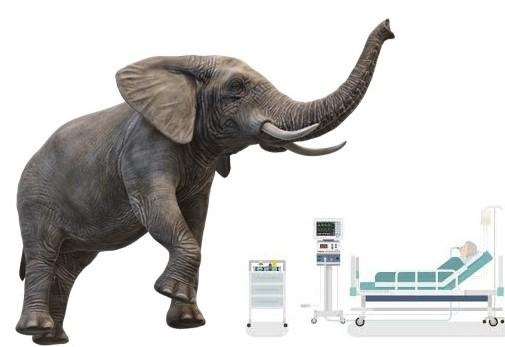
Ventilator Induced Lung Injury, the unseen elephant in the room

Those are traumas that happen after placing the patient on the ventilator and the only way to avoid them is to avoid mechanical ventilation which of course can’t always be done.

Those can cause worsening hypoxemia that can prolong mechanical ventilation, lead to multi-system organ dysfunction, and increase mortality.

So the best strategy is to try to avoid them, rapidly diagnose them, and correct what we can.

Our understanding of VILI have spiked over the last two decade. Since the positive results of the low vs high tidal volume trial in ARDS in the beginning of the century, most clinicians start to pay attention to low tidal volume and limiting plateau pressure but is that enough? Is that the answer?

So how are we doing with such understanding, any better? Unfortunately not too good, we can do better, need more work to be done, we owe it to our patients so let’s dig in this very complex problem.

What is VILI:

There are so many types and names we should all be familiar with (Volutrauma, Barotrauma, Biotrauma, Atelectrauma, Shear injury, Diaphragm myotrauma, Oxygen toxicity, SILI, Capillary endothelial injury, etc).

During mechanical ventilation, the lung is under continuous forces during inspiration and expiration, mainly: Stress, Strain, and the frequency of such forces. To be fair, those forces are not solely from the ventilator, but the patient himself play a big role.

Because of the marked heterogeneity of our lung units both in health and disease, different areas of the ventilated lungs are under different stresses (forces per unit area, represented by Trans-pulmonary pressure, i.e. the inside alveolar pressure minus the opposing outside pressure represented by pleural pressure), strain (the dynamic change in shape and deformation of the alveoli).

There has been a debate over the years of what is injurious to the lung, pressures, or volume. The answer is probably both and more. The Mechanical power equation sums the forces in one:

Mechanical Power = VE x (Peak Pressure + PEEP + F/6) / 20

However this equation lacks some important components: the patient-ventilator interaction, trans-pulmonary pressures, dead space, the ventilation distribution to different lung regions.

To summarize the forces we need to pay attention to minute ventilation, respiratory rate, Delta inspiratory pressures, PEEP, tidal volume, trans-pulmonary pressure, patient-ventilator asynchronies, FRC, dead space, and ventilation distribution.

Risks for VILI:

So who develops VILI? We know there are many risks for developing VILI and SILI (self-induced lung injury), but the true incidence and prevalence of VILI is unknown, majorly because of uncertainty in diagnosis.

The terms VALI (ventilator associated lung injury) or VAE (ventilator associated events) were developed based on worsening oxygenation and the need to increase FiO2 and PEEP because of the uncertainty in diagnosis and are very nonspecific and don’t solve the problem.

So why some patients develop VILI, and some don’t? Is it genetic phenotype? The answer is unknown similar analogy are why not all smokers develop COPD or lung cancer.

Diagnosis of VILI:

Unfortunately in clinical practice, it is very difficult to diagnose, with the exception of Barotrauma in the form of pneumothorax, pneumomediastinum which are easily diagnosed radiologically.

Clinical diagnosis remains the only available method and to rule out different other etiologies. Worsening oxygenation, new radiographic evidence of lung infiltrates and respiratory mechanics are usually attributed to infectious Ventilator Associated Pneumonia (VAP) or cardiogenic pulmonary edema leading to the wrong diagnosis, unnecessary antimicrobials and diuretics and most importantly not recognizing the true problem.

No labs or serological markers are currently available for everyday practice for early diagnosis of VILI. There are ample research using different biomarkers in animals, but they remain under investigation and limited to research labs.

Monitoring respiratory mechanics and waveforms can tell us early that there is a new problem but can’t diagnose if the new problem is VILI or just worsening of the initial etiology that landed the patient on the ventilator or other problems (TRALI, VAP, DAH, Pulmonary edema, etc)

What can we do at the bedside?

Obviously, we first need to prevent VILI.  “Prevention is better than cure”

In addition to monitoring and minimizing all the factors included in the equation of mechanical power, there are additional steps that can be taken into consideration.

Monitoring the trans-pulmonary pressure is very important to avoid the excess stress on the alveolar unit both at inspiration and expiration.

Optimizing the PEEP is also crucial according to physiological parameters not based on oxygenation or a table with the one hat fits all concept. Taking in consideration that not one level of PEEP regardless high or low is optimal to all alveolar units in both lungs that have their own opening and closing pressures and under different trans-pulmonary pressures stress.

Optimize and prevent patient-ventilator desynchronies as much as possible, acknowledging the thin line between spontaneous breathing benefits and the possible benefits of muscle paralytics and diaphragmatic dysfunction.

Baby lung or open lung approach: another controversial topic but points to consider are the baby lung is a functional unit and is not anatomical and is not equivalent to 6 ml/kg IBW (which is the normal tidal volume of a healthy person and could be still excessive in lung injury). The open lung approach concentrates on increasing the FRC of the lung through recruitment, different ventilator modes but also carry the risk of excessive volumes and pressures.

Prone position is one of the simple maneuvers that improves the lung inhomogeneity, that improves oxygenations, outcomes and probably reduces VILI but is still underutilized in ARDS, however its use during the COVID-19 pandemic has markedly increased for intubated and non-intubated patients.

Technologies that monitor lung volumes, e.g. CT scans are available but carries the risk of radiation exposure and transporting unstable patients to radiology suits (though mobile units are available). Electrical Impedance Tomography (EIT) is a technology that can be used continuously at the bedside to monitor lung volumes and assess over and under distension in real time helping the clinicians adjust the volumes, pressures, and position of the patient.

Secondly, we need to improve our diagnosis and adjust our strategies of ventilation if VILI is occurring, otherwise we become “The blind leading the blind”

The deficit in diagnosis is a major obstacle. Education is absolute necessity, research on biomarkers of VIL is crucial (similar to troponins as a marker of myocardial injury, and lactic acid as a hypoperfusion marker in shock states). We need reliably sensitive and specific markers (serologic or based on breath analysis), fast, not too expensive so can be incorporated at the bedside.

The future:

With the advance in technology, education, research, artificial intelligence, I am extremely hopeful that we will do better eventually specifically in the topic of VILI and mechanical ventilation in general.

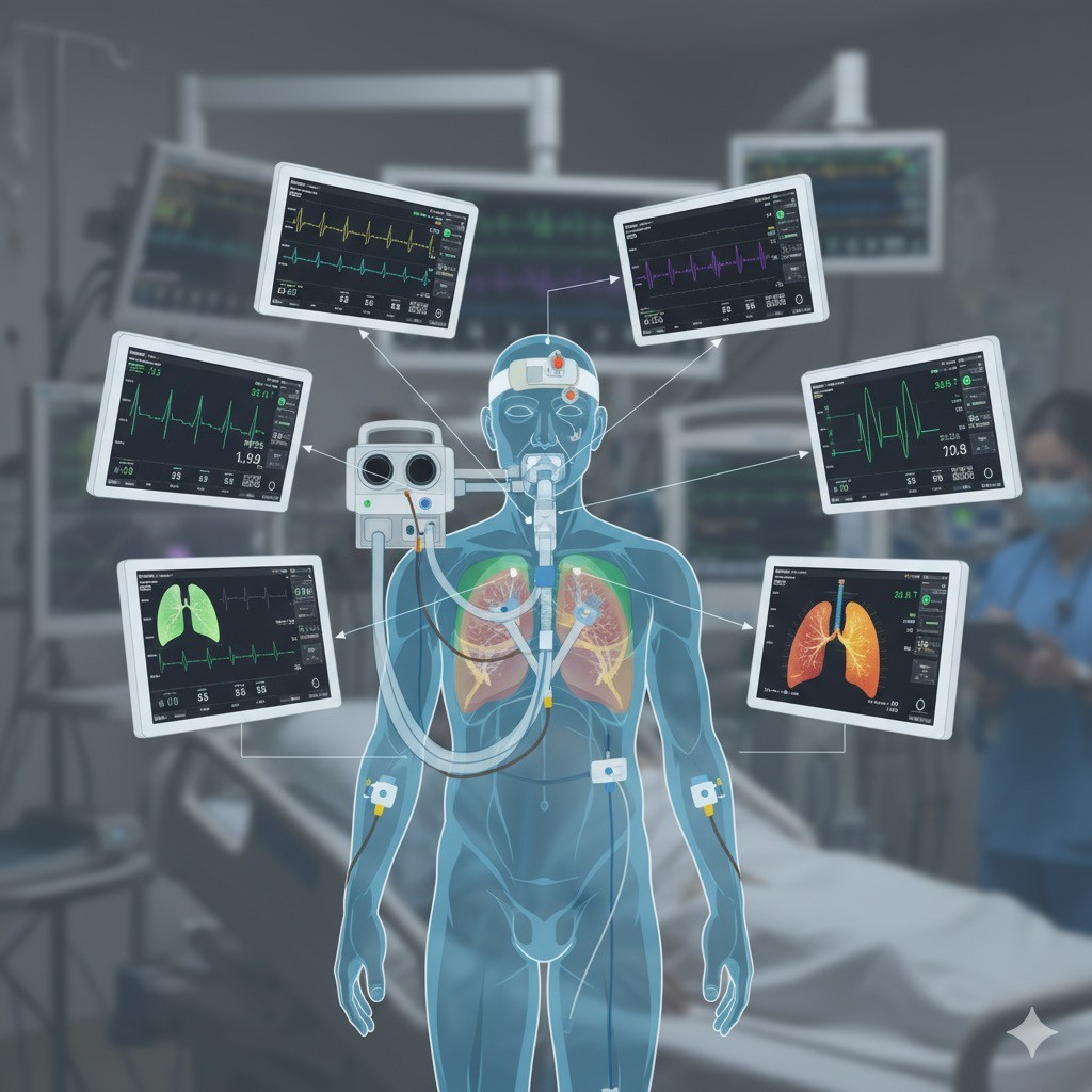
I can see alert systems all incorporated in one place probably within the ventilator that incorporates different measures and maybe analyze the expiratory gas from the patient recognizing early VILI and automatically correct the issues or gives suggestions to the bedside clinicians.

To summarize:

Personalized ventilation to each patient

Spend the time at bedside

More education and improve enthusiasm

More research

Uniting forces from all who have stake in mechanical ventilation (clinicians, societies, universities, manufacturers)

New technologies and artificial intelligence
The Revolution Begins (The 6 ml/kg)

In 2000, I was an internal medicine resident highly fascinated with yet poorly educated about the field of critical care medicine specifically the science of mechanical ventilation. When the ARMA trial of low tidal volume 6 vs 12 ml/kg IBW came out that year, I thought why 12? Did we even use 12 ml/kg even then?

The joke between us at the time was, any patient goes on the ventilator, the settings are AC (I never use that horrible term unless I am talking about air conditioning), tidal volume 500, respiratory rate of 15 and PEEP 5 regardless. So, for an average patient with IBW 70 kg, that is equivalent to 7.1 ml/kg, little higher for lower IBW and little lower for higher IBW. I cannot believe we used 840 ml for an average person to start with.

For the last 21 years, I have been arguing that it is not the tidal volume only that injures or add injury to the injured lung, it is the pressures applied to the alveoli that causes the stress and strain on those alveoli. Not only the plateau pressure or the driving pressure alone but the sum of forces (trans-pulmonary pressure) from inside (plateau pressure) and outside (pleural pressure) as estimated by an esophageal balloon.

I just refuse the one size hat fits all concept, just does not make any physiological sense.

The 6 ml/kg could be injurious to a lung that has > 50% of alveoli collapsed, and 10 ml/kg could be ok for a lung that has most of its alveoli open via the “open lung approach”.

Heretic and ignorant was called to unbelieve or disregard the ARDS network and their guidelines that were adopted by every society and hospital. Though in every case of mechanical ventilation, I try to use the lowest tidal volume, lowest driving pressure and lowest plateau pressure I can achieve. In difficult cases, I indeed use the esophageal balloon manometry to calculate the total respiratory compliance and its two components: lung and chest wall, and guide my pressures applied (driving pressures and PEEP) through monitoring the end inspiratory and end expiratory trans-pulmonary pressures even in the non- conventional modes like APRV and during prone position.

I am also still waiting for the Electrical Impedance Tomography (EIT) to be commercially more available as it will give us a whole new insight in this issue and of how we apply mechanical ventilation.

But I was not alone, highly respected great minds in mechanical ventilation (Tobin, Amato, Gattinoni, just to name a few) have argued that it is the pressure whether plateau pressure, driving pressure that is injurious. The eternal fights between the most famous and intelligent minds in mechanical ventilation filled the literature of whether it is the volume or the pressure, with evidence to support their claims. I would add that it is probably both.

We also do not talk much about the asynchronies that happen a lot especially in the low tidal volume strategy that are injurious by themselves.

A recent meta-analysis of five randomized trials was published this month of July 2021 have shown that indeed it is the driving pressure more than the tidal volume that worsens mortality through ventilator induced lung injury. An editorial for that study by Dr. Tobin (one of the people you should listen to about mechanical ventilation) titled “The Dethroning of 6 ml per kg as the “go-to” Setting in ARDS” made me jump to write this in support of his and others who are fighting the good fight of ventilator induced lung injury.

I think the time has come to treat every patient on a physiologic basis and parameters not like cars were the gas tank gets filled with a fixed certain amount of gas.

To summarize, I want to be clear, I am not advocating for high tidal volumes but advocating for the lowest safest tidal volume, driving pressures, plateau pressures, transpulmonary pressures possible.

Yet the hard reality is our lungs and respiratory system in general are very heterogenous, and ventilator lung injury to some extent cannot be totally prevented as different areas of the lungs are not subjected to the same tidal volumes, driving pressures, plateau and transpulmonary pressures. Those are the sum of both lungs. Unless we can ventilate each lobe, each segment, each subsegment separately.

Not to leave at a pessimistic view, I am very hopeful that our understanding, monitoring and treatment techniques will continue to grow and improve.

Share on Social Media
Follow us on Social Media